 Case Report
Use of Naso-Melo-Labial Flap of Advancement and Rotation in Canine Bite Injury. About 1 Case.
Marco Antonio Aldaco Aguirre1* and Edith Yamilet Mendiola Castillo2
1Otolaryngology and Facial Surgery, National Autonomous University of Mexico/Mexican Council of Otolaryngology and Head and Neck Surgery, Mexico
2University of the Northeast, Tampico, Tamaulipas, Mexico
Dr. Marco Antonio Aldaco Aguirre, Otolaryngology and Facial Surgery, National Autonomous University of Mexico/Mexican Council of Otolaryngology and Head and Neck Surgery, Mexico.
Received Date:July 18, 2022; Published Date: September 14, 2022
Abstract
A 40-year-old male with a past medical history of acute mental status (mixed neurosis), suffered from a canine bite (pitbull) in the nasal region.
The patient is presented to the emergency room in a second-level private hospital to comence reconstructive treatment. He showed a bite of less than
2hrs of evolution, with loss of substance partially of the back, right lateral wall, ipsilateral nasal wing, with skin involvement, subcutaneous cellular
tissue, right inferior and superior wing cartilage.
The wound was washed, debrided and prophylaxis was performed, followed by the planning of a combined local flap, this being an advancement
and rotation nasomelolabial flap, as well as harvesting and application of autologous graft of the right atrial concha. This procedure was done in a
single surgical intervention. The aesthetic and functional results were classified as favorable. The flap technique is discussed, depending on various
forms of reconstruction.
Keywords:Nasogenien flap; Nasolabial flap; Frontonasal flap; Wound; Substance loss; Total skin graft; Wing of the nose; Nasal pyramid
Introduction
The nose reconstruction is a very broad topic, covering innumerable situations and variety of repair techniques, which depend on: the etiology (whether tumor, traumatic or congenital), the characteristics of the patient (children or elderly, men or women, healthy people, smokers or with chronic-degenerative diseases), the topography (upper part of the nasal edge, tip, back or nasal wing), the extent and depth of the lesion (loss of subtotal, total substance or mutilation).
We must consider the definition of a flap, referring to healthy skin and tissue that moves and partially separates to cover nearby bloody areas. This involves skin, fat, muscle, or a combination thereof, which in the pedicle ensures vascular input.
In this special area, the aesthetic and functional requirement is very high and requires reconstructions close to normal, which is possible, thanks to technical advances and the way in which they are employed. Recently, great expectation has been generated regarding the flap technique, a procedure that has an increasing use in coverage of bloody areas, traumatic wounds in the nasal pyramid, due to the effects on aesthetics and function. In addition to having advantages of satisfactory results and a short-term recovery, as well as favorable cost benefit with outpatient management. So today, the vast majority of reconstructive surgeons opt for this surgical modality that offers quite favorable results, avoiding techniques that require several surgical procedures, without interfering vastly with the respiratory function of the nasal cavity.
Within the etiology of lesions in the nasal pyramid, there are injuries due to aggression of the external environment (burns, trauma wounds with blunt and cutting object, chromator workers and inhaled drugs) or internal processes (benign or malignant neoplasms, granulomatous disease and, mycosis), which can generate loss of substance in the different regions. Therefore, the optimal reconstruction technique depends on the location, extent, depth, and experience of the surgeon.
The types of flaps can be rotation (on its axis), advancement (in a straight line forward) and transposition (in a straight line to the sides of the flap), on island, tubular. In addition, due to its irrigation, the flap can be: ramdomized (when there a non-extistential source of vascularization and is only composed of branches of major blood vessesls) or axial (when it has in its pedicle a recognized main arterial vessel), according to the donor area there are frontal, melolabial, cervices-facial, etc.
We present this case in order to provide reconstructive plastic surgeons, otolaryngologist head and neck surgeons a viable surgical alternative to reconstruct injuries with loss of substance focused on the nasal pyramid, which nowadays occur very frequently. This technique aims to get closer to the natural aesthetic and functional parameters of every single patient.
Clinical Case
A 40-year-old male with a past medical history of acute mental status (mixed neurosis), suffered from a canine bite (pitbull) in the nasal region. 30 minutes after, the wound was profoundly cleansed by a board-certified doctor.
The patient is presented to the emergency room in a secondlevel private hospital to comence reconstructive treatment.
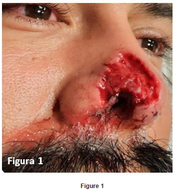
He showed a bite of less than 2hrs of evolution, with loss of substance partially of the back, right lateral wall, ipsilateral nasal wing, with skin involvement, subcutaneous cellular tissue, right inferior and superior wing cartilage (Figure1).

Initial treatment included prophylactic antibiotics in the view of the fact that the canine bite is potentially contaminated
Given the case, double scheme with cephalosporins and lincosamides incitated. As well as ceftriaxone 1gr iv every 12hrs for 10 days and clindamycin treatment of 600mg iv every 8hrs for 5 days given the resistance to beta-lactams in our environment. In order to avoid tetanus and rabies, anti-rabies and anti-tetanus immunization was appointed. In the emergency room a complete medical history, as well as preoperative laboratory examinations, SARS-Cov2 antigen test (negative), exhaustive mechanical washing (decontamination cure) are performed and at 24 hours after prophylaxis the operative technique chosen for this case was performed.
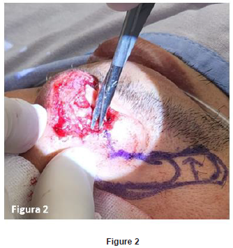
Under local anesthesia and sedation prior asepsis and antisepsis of the region, placement of sterile fields, followed by marking of the flap of choice with a diameter of 30mm in length x 15mm in width (Figure 2), this flap being naso-melolabial of advancement and rotation. The bleeding area to be treated has 25mm of caudal cephalic length x 20mm of width, with a pyramidal shape of lower base on the right-wing edge, infiltrates with articaine plus epinephrine 1/50,000 iu (medicaine) in the surgical areas to be treated, cartilaginous ingestion of right atrial shell (donor area) is obtained to place it at the site of the lower and upper wing cartilage (receiving area) fixing it with prolene 5-0 (Figure 3). To obtain the flap, an incision of skin and subcutaneous cellular tissue using scalpel number 15 Miltex in the marking line of the surgical plan is necessary. Performing subcutaneous dissection with stevens scissors of 14cm double edge blunt tip to continue with electronic scalpel with colored tip, hemostasis is corroborated being conservative in cauterization to preserve the vascular contribution greatly. This incision is continued to the wing groove of the affected area, then we proceed to the dissection of the planned flap including the nasal lateral wall, perform advance and block rotation of the dissected tissue giving total coverage to the bleeding area, seeking a minimum tension in the edges of coping and skin suture. Closure of surgical edges is performed by planes using vicryl 3-0 in internal plane inverted points and dermalon 5-0 for skin closure with simple points (Figure 4), internal nasal splint is placed with Doyle type tamponade with ventilation tube which allows us postoperative nasal permeability avoiding at the same time scar stenosis of the affected nostril. Antibiotic ointment is applied on the edge of the suture and placement of sterile gauze dressing. Surgical act is terminated without incidents or accidents.


Discusion
We observe that prompt intervention in this type of case within 8 hours of the injury, with exhaustive mechanical lavage and starting prophylactic coverage, anti-rabies, and anti-tetanus immunizations within 24 hours, gives us the guideline to perform a definitive surgical coverage procedure for the bleeding area with a naso-melo-labial advancement and rotation flap supported by the verification of good vascularity and cleanliness of the injured área.
Discussion
In the immediate postoperative period, the surgical margins were practically without tension, with minimal right lateralization of the columella, discreet edema, adequate capillary filling of the flap. In its evolution at 7 days, external sutures were removed and the endonasal splint was removed at 10 days, without presenting nostril stenosis, daily dressings were performed every 12 hours with antiseptic solution (Mycrodacin) and surgical soap.
After two months of evolution, healing has been favorable, with a satisfactory cosmetic and functional result, together with an improvement in his self-esteem and quality of life (Figure 5). It is expected that at the end of the scar fibroplasia process it will be almost imperceptible [1-9].

Conclusion
Timely intervention, even in the case of a canine bite, is essential to make the decision to initiate decisive treatment in this type of case. Choosing the donor area adjacent to the recipient area increases the chances of success in a single surgical time. With a positive cost-benefit balance for our patient and a short-term recovery.
Acknowledgement
None.
Conflict of Interest
No conflict of interest.
References
- Quintana Díaz JC, Villarreal Corvo N (2017) Reconstrucción de defecto facial causado por mordedura canina. MediMay.
- Pérez Cánovas C (2019) Mordeduras y picaduras de animales. En: Sociedad Española de Urgencias de Pediatria. Protocolos diagnósticos y terapéuticos en urgencias de Pediatrí (3rd edn.), Madrid: SEUP, Spain.
- Young WG, Worsham MJ, Joseph CL, Divine GW, Jones LR, et al. (2014) Incidence of keloid and risk factors following head and neck surgery. JAMA Facial Plast Surg 16(5): 379-350.
- Chávez Serna E, Andrade Delgado L, Martinez Wagner R, Altamirano Arcos C, Espino Gaucin I, et al. (2019) Experience in the management of acute wounds by dog bite in a hospital of third level of plastic and reconstructive surgery in Mexico. Cir Cir 87(5): 528-539.
- Diaz Fernández JM, Mestre Cabello J (2017) Reconstrucción labial por mordedura canina en una anciana. MEDISAN.
- Miranda Rius J, Brunet LLobet Ll, Lahor Soler E, Mendieta C (2014) An unexpected presentation of a traumatic wound on the lower lip: a case report. J Medical Case Reports 8: 298.
- Khandelwal P, Hajira N, Dubey S (2015) Management of maxillofacial injuries in humans due to animal bites and mauling: A report of three cases. Niger Postgrad Med J 22(4): 241-244.
- Venkatesh KL, Bhaskan A, Vepakkomma D (2017) Study of morbidity of pediatric dog bites: single center experience. Int Surg J 4(1): 185-188.
- Gopinath AL, Reyazulla MA, Ajay Kumar N, Kadanakuppe S (2015) A stich in time saves nine: Primary closure in facial dog bite injuries. A case series. Open J Dent Oral Med 3(1): 7.
-
Marco Antonio Aldaco Aguirre* and Edith Yamilet Mendiola Castillo. Use of Naso-Melo-Labial Flap of Advancement and Rotation in Canine Bite Injury. About 1 Case.. On J Otolaryngol & Rhinol. 5(5): 2022. OJOR.MS.ID.000621.
-
Nasal wing, Reconstructive treatment, Nasal region, Nose reconstruction, Nasal edge, Nasal cavity, Internal nasal splint, Naso-melolabial, Local anesthesia
-

This work is licensed under a Creative Commons Attribution-NonCommercial 4.0 International License.


